Log in and enrol
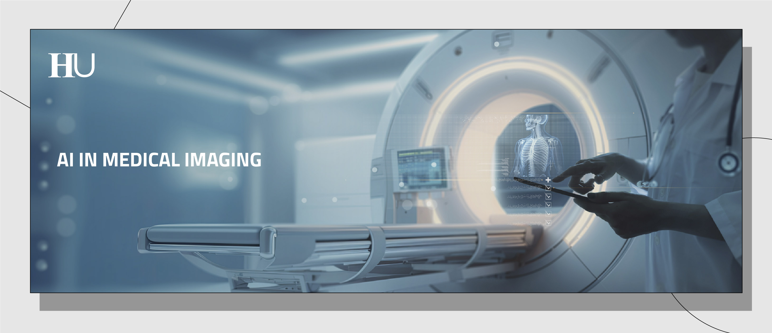
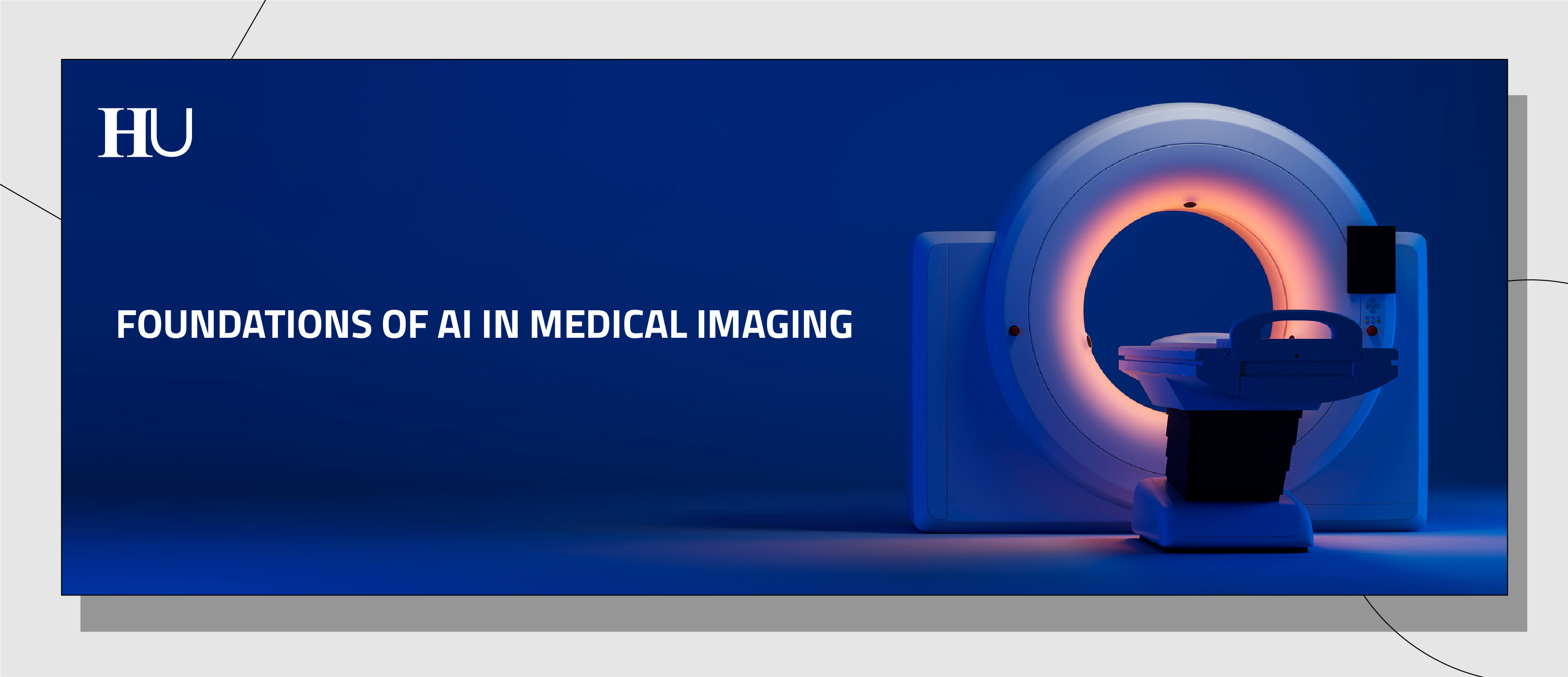
AI in Medical Imaging
AI in Medical Imaging
Course description
How is AI changing the way we see disease and the way hospitals turn images into decisions?
This MOOC explores AI in medical imaging as a bridge between pixels and patient care, showing how data move from scanners to clinical insight without losing safety, trust, or disrupting everyday clinical workflows.
As you progress through the three weeks, you’ll explore how AI learns to read X-ray, CT, MRI, PET/SPECT and digital pathology, and how imaging data become reliable signals for clinical decisions. We’ll also address what it takes to deploy these tools in real hospitals: integration into everyday workflows, monitoring drift, reducing bias and protecting privacy through collaboration at scale.
Through a blend of video lectures, interviews, quizzes, case studies, and discussions, you’ll also gain practical insights into the implications of AI in medical imaging. By the end of this course, you’ll be equipped to critically assess what it takes to make imaging AI work in real hospitals, understanding its potential to support diagnosis and clinical workflows while meeting safety, equity, and regulatory responsibilities.
Total workload of the course: 12 hours
This MOOC was produced as part of the Edvance project – Digital Education Hub per la Cultura Digitale Avanzata. The project is funded by the European Union – Next Generation EU, Component 1, Investment 3.4 “Didattica e competenze universitarie avanzate".


Intended Learning Outcomes
By the end of this course, here are some things you’ll be able to do:
- Describe How Medical Images Become AI-Ready Data: You’ll be able to explain how medical images become usable inputs for AI through DICOM, and why the surrounding clinical systems (PACS/RIS/EHR) and data silos shape what is feasible in real practice.
ESCO: medical imaging technology - Compare AI Approaches in Imaging: You’ll be able to describe how radiomics and deep learning extract clinically meaningful patterns from images, and explain the evolution from CNNs to Transformers across radiology, nuclear medicine, and digital pathology (including WSI).
ESCO: utilise machine learning - Understand Clinical Integration and Adoption Barriers: You’ll be able to identify the main barriers to AI adoption in clinical settings, including workflow disruption, interoperability constraints, and usability factors that influence adoption, and explain why accuracy alone does not guarantee clinical sustainability.
ESCO: make clinical decisions - Identify Regulatory Responsibilities Under the EU AI Act: You’ll be able to recognize the operational implications of the EU AI Act for healthcare organizations, including governance expectations and the responsibilities tied to deploying high-risk AI systems in routine care.
ESCO: medical device regulations - Detect Real-World Risks and Equity: You’ll be able to detect post-deployment threats such as performance drift and domain shift, and recognize demographic bias that can undermine fairness and trust in clinical decision-making.
ESCO: data quality assessment - Recognize New Paradigms for Scale and Collaboration: You’ll be able to describe foundation models as a response to fragmented point solutions, and explain how federated learning enables multi-institution training while keeping sensitive data local.
ESCO: protect personal data and privacy
Prerequisites
Don't Sweat It!
This is an introductory course—no need to be an engineer or an AI expert. Though a basic familiarity with medical terminology and clinical workflows is helpful.
If you're a student in a single-cycle Medicine and Surgery program (from the third year onward), a third-year Bachelor's program in nursing and healthcare technology, or a Master's degree in the health sciences, you'll be just fine!
Our goal is to make AI in medical imaging understandable and clinically grounded: how pixels become signals, how models are evaluated, and what it takes to deploy them safely under real-world constraints—regulation included.
We’re here to make AI easy to understand and show you how it applies to medical imaging. If you’re curious about diving deeper into the tech side, don’t worry—this portal has plenty of advanced AI courses. Don’t be shy, explore!
Activities
Video lectures and expert guest talks will guide you through the main topics of the course, alongside additional readings with reflective questions to support your learning. Along the way, you'll encounter short, ungraded quizzes and challenges designed to check your understanding, encourage hands-on practice, and spark discussions in the Forum (a.k.a. The Black Box Café). Be sure to complete the mandatory milestone quiz at the end of each week—passing these is required to earn your certificate.
Meet The Black Box Café!
This is The Black Box Café, our Discussion Forum.
You’ll meet The Black Box Café again as you go through the course. It’s not mandatory to engage with it. Neither we nor any AI system will be monitoring it. This is just a space for you to connect with your fellow humans during your learning journey.
Think of The Black Box Café as the slow food version of social media — less noise, more substance.
Section outline
-
-
We’ll kick off by building a shared foundation on how medical images are created, stored, and interpreted in clinical practice—from X-ray, CT, and MRI to PET/SPECT and digital pathology. Along the way, we’ll see how AI is learning to “read” these images through standards like DICOM, approaches like radiomics and deep learning, and why all of this matters for diagnosis and research.
-
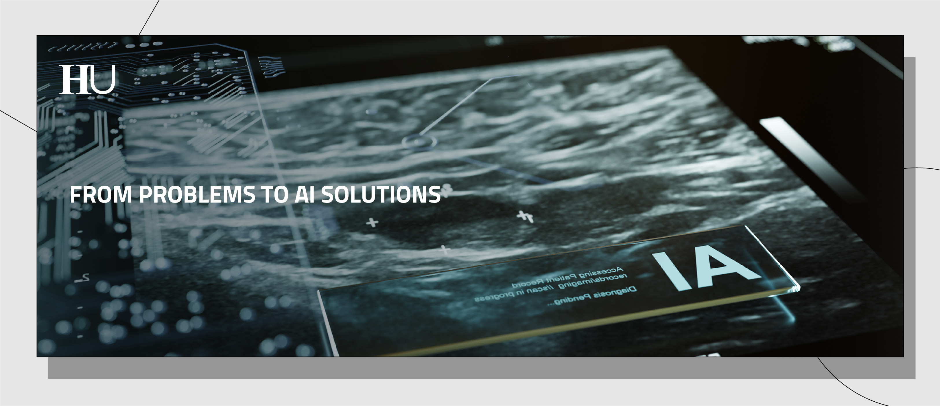
How can AI help clinical teams keep up with growing imaging complexity— without compromising accuracy or safety? In this module, we’ll explore how AI is solving real-world clinical challenges in imaging interpretation and operational efficiency, showing how these tools can support faster workflows, more consistent assessments, and more informed decisions in everyday practice. Along the way, you’ll hear from double interviews that bring together both the clinical perspective and the technical perspective, helping you connect everyday clinical needs with the technical foundations behind these tools.
-
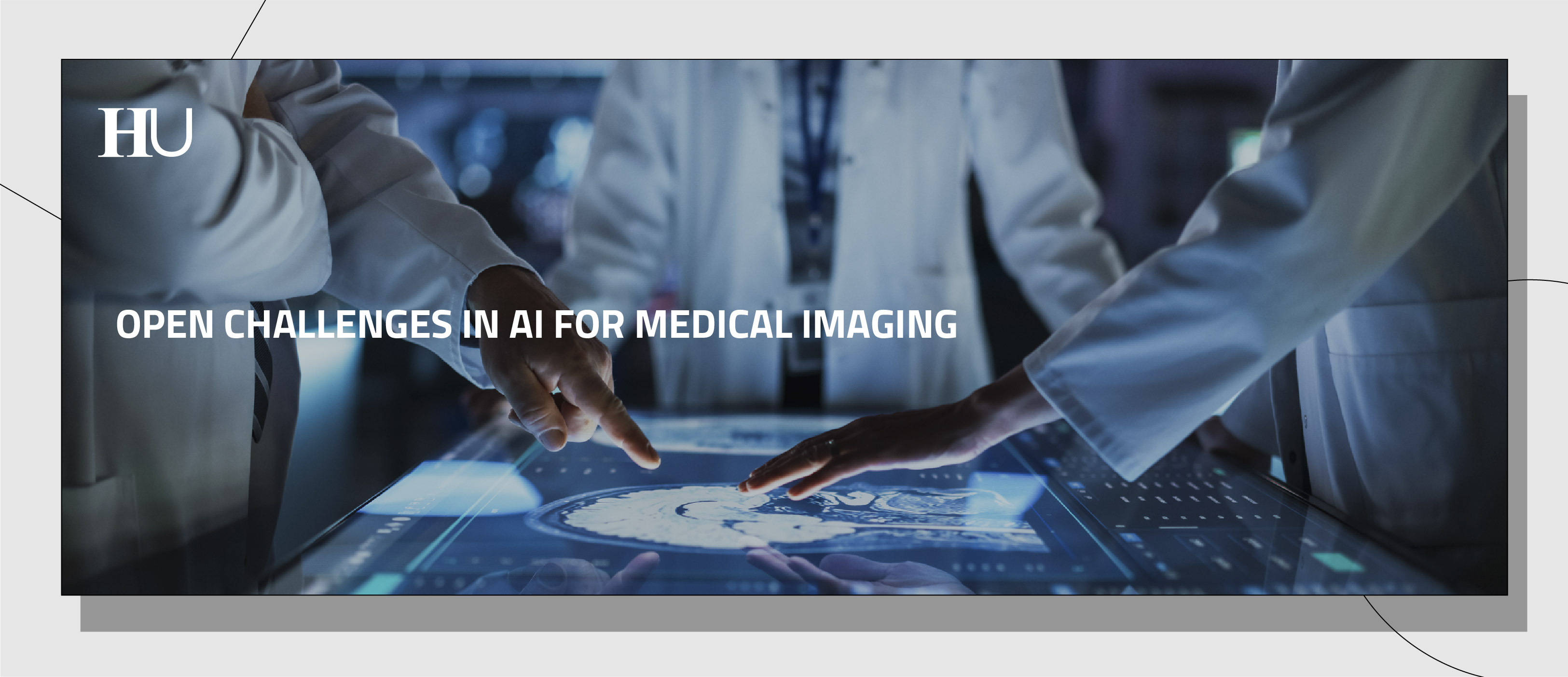
This week we shift from “does the model work?” to a more practical question: what happens when AI tools start piling up in the workflow without a clear strategy? We’ll unpack why clinical AI often becomes an integration and lifecycle challenge—plugging results into PACS/RIS/EHR, fitting real timing constraints, and staying usable for clinicians. From there, you’ll get a guided view of what’s changing at scale, from task-specific tools to newer foundation-model approaches, and why privacy and data silos make collaboration across hospitals harder than it sounds.
We’ll close by connecting these ideas to governance and responsibilities in real deployment—so AI can support care without adding new burden to the people delivering it. -
-
Video transcripts Folder
-
Assessment
Your final grade for the course will be based on the results of your answers to the assessed quizzes. You have an unlimited number of attempts at each quiz, but you must wait 15 minutes before you can try again. You will have successfully completed the course if you score 60% (or higher) in each one of the assessed quizzes. The maximum score possible for each quiz is given at the beginning of the quiz. You can view your score in the quiz on your last attempt or on the 'Grades' page.
Certificate
You can achieve a certificate in the form of an Open Badge for this course, if you reach at least 60% of the total score in each one of the assessed quizzes and fill in the final survey.
Once you have completed the required tasks, you will be able to access ‘Get the Open Badge’ and start issuing the badge. Instructions on how to access the badge will be sent to your e-mail address.
The Badge does not confer any academic credit, grade or degree.
Information about fees and access to materials
The course is delivered in online mode and is available free of charge.
Course faculty
Prof. Letterio Salvatore Politi
Full Professor of Neuroradiology at Humanitas University
Prof. Letterio Salvatore Politi is a Full Professor of Neuroradiology at Humanitas University. With over 20 years of clinical experience, his research focuses on advanced MRI methods for non-invasive quantification of disease burden, aiming to define disease natural history and outcome measures.
He is also the Co-Director of the Residency Program in Radiodiagnostics at Humanitas, collaborating with Prof. Marco Francone. At IRCCS Humanitas Research Hospital, Prof. Politi directs the Neuroradiology Department, focusing on brain tumors, neurodegenerative diseases, and neuromuscular disorders. His research group works on artificial intelligence applied to radiology and advanced MRI methods.
Prof. Riccardo Levi
Assistant Professor of Biomedical Engineering at Humanitas University
Prof. Riccardo Levi is an Assistant Professor at Humanitas University, specializing in biomedical engineering with a focus on neuroradiology. He earned his PhD in biomedical engineering and is currently a researcher at the Department of Biomedical Sciences. Prof. Levi's research primarily concentrates on developing deep learning algorithms for neuroradiology applications. He serves on the scientific editorial board of the journal European Radiology (Neuro section) and is a member of the European Society of Medical Imaging and Informatics Young Club.
Prof. Laura Evangelista
Extraordinary Professor of Diagnostic Imaging and Radiotherapy at Humanitas University
Prof. Laura Evangelista is Extraordinary Professor of Diagnostic Imaging and Radiotherapy at Humanitas University in Milan, Italy, and Head of the Nuclear Medicine Group across Humanitas Hospitals (including Rozzano, Bergamo, and Catania). She trained in Nuclear Medicine at the Università degli Studi di Napoli “Federico II” and completed a PhD in Molecular Imaging, further enhancing her expertise with international fellowships and advanced training. Prof. Evangelista’s clinical and research work focuses on nuclear medicine and oncological imaging, particularly the use of innovative radio‑pharmaceuticals for diagnosis and therapy in prostate, breast, and lung cancers. She leads and contributes to multicenter studies on PSMA PET/CT and other molecular imaging modalities to improve cancer staging and treatment monitoring. Prof. Evangelista has published extensively in high‑impact journals, authored numerous book chapters, and serves as Editor‑in‑Chief of Clinical and Translational Imaging, the official journal of the Italian Association of Nuclear Medicine. She is actively involved in professional societies and initiatives promoting translational imaging research and women’s leadership in nuclear medicine. Her academic role includes teaching across medical and allied health degree programs at Humanitas University.
Prof. Giuseppe Jurman
Associate Professor of Biomedical Engineering at Humanitas University
Prof. Giuseppe Jurman is an Associate Professor at Humanitas University, with data science as his main research interest. His focus is applying mathematical and computational methods to life sciences, in particular the development of predictive models based on artificial intelligence. Prof. Jurman leads the Data Science in Health (DSH) Unit at Fondazione Bruno Kessler (FBK) in Trento, Italy, where his research focuses on machine learning, network analysis, and their applications in healthcare. He has published extensively in these areas, contributing significantly to the intersection of data science and biomedical research.
In January 2025, Prof. Jurman was appointed as an Associate Professor at Humanitas University, where he continues to advance research in data science and its applications in health.
Prof. Salvatore Piscuoglio
Associate Professor of Genetics at Humanitas University
Prof. Salvatore Piscuoglio is Associate Professor of Genetics at Humanitas University and Group Leader of the Precision Medicine Research Lab at Humanitas Research Hospital in Milan, Italy, where he leads multidisciplinary efforts to advance cancer precision oncology. He earned his degree in Medical Biotechnology from the University of Naples “Federico II” and completed his PhD in Genetics at the University of Basel, Switzerland, followed by postdoctoral training at the Memorial Sloan Kettering Cancer Center in New York. His research focuses on the identification of novel cancer genes, predictive biomarkers of therapy response or resistance, and discovery of new therapeutic targets through integrated multi‑omics profiling and computational approaches. Prof. Piscuoglio has held leadership roles at the Institute of Medical Genetics and Pathology and Department of Biomedicine at the University Hospital Basel prior to joining Humanitas. His group employs patient‑derived models and ex‑vivo drug profiling to bridge genomic insights with translational applications. He contributes to national and international research collaborations aimed at personalized cancer treatment strategies. Prof. Piscuoglio is active in scientific dissemination and regularly participates in conferences focused on precision oncology and biomarker discovery. His work supports the development of innovative approaches to improve cancer diagnosis, prognosis, and therapy.
Prof. Andrea Laghi
Professor of Radiology at Humanitas University and Director of the Department of Diagnostic Imaging at Humanitas Research Hospital
Prof. Andrea Laghi is Professor of Radiology at Humanitas University in Milan and Director of the Department of Diagnostic Imaging at Humanitas Research Hospital. He trained in radiology at Università La Sapienza – Rome and has extensive clinical and research experience in abdominal and oncologic imaging, including the application of advanced imaging techniques and artificial intelligence in diagnostic radiology. Prof. Laghi is also a member of the Consiglio Superiore di Sanità (Italian National Health Council) and has served in leadership roles in major scientific societies such as the European Society of Gastrointestinal and Abdominal Radiology (ESGAR), where he was President, and the European Society of Oncologic Imaging (ESOI). His research interests encompass abdominal CT and MRI, hybrid imaging, and AI‑enhanced diagnostic workflows, with numerous peer‑reviewed publications and invited presentations at international conferences. He contributes to European and national research initiatives aimed at integrating advanced imaging and computational methods into clinical practice. Prof. Laghi teaches and mentors students and residents in radiology and participates in interdisciplinary projects that bridge imaging, data science, and personalized medicine. His work supports the advancement of diagnostic precision and the clinical impact of radiological science.
Prof. Gian Marco Conte
Assistant Professor of Radiology in the Department of Radiology at Mayo Clinic in Rochester, Minnesota
Prof. Gian Marco Conte, M.D., Ph.D. is Senior Associate Consultant, AI Scientist, and Assistant Professor of Radiology in the Department of Radiology at Mayo Clinic in Rochester, Minnesota. He earned both his medical degree and Ph.D. from Vita‑Salute San Raffaele University in Milan, Italy, where his early research focused on advanced perfusion MRI in neuroimaging. At Mayo Clinic, Dr. Conte’s work bridges clinical radiology and artificial intelligence, emphasizing the integration of machine learning techniques into medical imaging to enhance diagnostic accuracy and translational practice. He has completed postdoctoral research in AI and medical imaging, with applications in neuroimaging and neuro‑oncology. Dr. Conte serves on the editorial boards of Radiology: Artificial Intelligence and the Journal of Imaging Informatics in Medicine, contributing to peer‑reviewed scientific discourse. He is actively involved in professional committees dedicated to advancing imaging informatics and reproducible AI research. His scientific contributions include work on deep learning models for tumor segmentation and diagnostic differentiation in MRI, reflecting his commitment to AI‑driven innovation in radiology. Dr. Conte’s research activities extend to benchmarking and collaborative challenges that promote fair and robust AI evaluation in healthcare.

Prof. Gianluca Cesare
Group Chief Information Officer at Humanitas Research and University Hospital Group
Gianluca Cesare is the Group Chief Information Officer at Humanitas Research and University Hospital Group, where he leads strategic planning and execution of digital health and information technology initiatives across the organization. He is a chartered engineer and completed a postgraduate Master’s in Supply Chain Management at Cranfield School of Management (UK). In his role at Humanitas, Cesare oversees the development and integration of health information systems aimed at enhancing clinical processes, data management, and patient‑centered services. Over more than a decade, he has played a pivotal role in major national digital health projects, including leading the Electronic Health Record (EHR) implementation for Regione Lombardia and initiatives in patient relationship management systems, establishing him as a key expert in digital health transformation in Italy. His work involves fostering innovation in healthcare IT, promoting interoperability, and leveraging data to support quality, safety, and efficiency in health services. Cesare collaborates with academic and industry partners and contributes to discussions on public‑private digital health strategies at national and international forums. Cesare is a member of several Advisory Boards among others EU - HealthCare Cybersecurity. He also serves as Adjunct Professor of ICT for Medicine at Humanitas University, teaching the intersection of information technology and healthcare. His professional experience spans healthcare informatics, consulting, and strategic IT leadership across sectors.
Dr. Walter Riviera
AI Engineer and Machine Learning specialist
Walter Riviera is a Computer engineer with over 10 years of experience, he previously served as Technical Director for AI at Intel, overseeing initiatives across Europe, Southeast Asia, and Africa. He is the author of more than nine scientific publications and was recognized by La Repubblica among Italy’s leading figures in artificial intelligence in 2024. He is also co-author of the book “L’Algoritmo dell’Uguaglianza”, featuring a foreword by Liliana Segre. Since 2025, he has been Chief AI Officer at AI Wonder, where he brings private AI solutions to Italian companies. He believes in the positive impact that responsible AI can have on society and is personally committed to raising awareness among both experts and non-experts through a theatre show he conceived and wrote.
Prof. Ciro Franzese
Associate Professor of Radiation Oncology at Humanitas University
Prof. Ciro Franzese, M.D. is Associate Professor of Radiation Oncology at Humanitas University and a specialist in Radiation Oncology and Radiosurgery at IRCCS Humanitas Research Hospital and Humanitas San Pio X in Milan, Italy. He graduated in Medicine from the Second University of Naples and completed his residency program in Radiation Oncology at the University of Florence, with additional research experience in the United Kingdom. Prof. Franzese is internationally recognized for his expertise in stereotactic body radiation therapy (SBRT) and stereotactic radiosurgery (SRS) for primary tumors and oligometastatic disease, particularly in genitourinary and other solid tumors. He has played leading roles in national and international clinical trials, he serves as Core Expert of the ESTRO SBRT Focus Group, and served as Coordinator of the AIRO Uro‑Oncology Study Group, contributing to advancements in radiotherapy practice and evidence‑based care. His research interests include advanced radiotherapy techniques, adaptive radiotherapy, and multidisciplinary treatment strategies. Prof. Franzese is actively involved in teaching and mentoring residents and fellows in radiation oncology, medical oncology, and diagnostic imaging. He has authored over 200 peer‑reviewed scientific papers and book chapters, reflecting his strong research profile. He also collaborates with professional societies such as SIURO and ISRS to disseminate best practices in radiotherapy worldwide. His work bridges clinical innovation, education, and research in cancer treatment.
Dr. Nicola Lambri
PostDoc and medical physics resident in the Department of Radiotherapy and Radiosurgery at IRCCS Humanitas Research Hospital
Nicola Lambri is a PostDoc and medical physics resident in the Department of Radiotherapy and Radiosurgery at IRCCS Humanitas Research Hospital in Rozzano, Milan, where he contributes to clinical and translational research in advanced radiotherapy. He is also affiliated with the Department of Biomedical Sciences at Humanitas University, participating in academic activities and research projects. His work focuses on developing and applying automated and machine learning‑based methods to streamline radiotherapy planning and segmentation workflows, with applications such as total marrow and lymphoid irradiation. Lambri’s contributions include co‑authorship of publications in international peer‑reviewed journals addressing automation and optimization in radiotherapy. He collaborates with multidisciplinary teams that integrate medical physics, clinical oncology, and computational approaches to improve treatment accuracy and efficiency. His research also engages with advanced imaging, artificial intelligence, and Lean Six Sigma methodologies to enhance clinical workflows. Lambri is active in presenting scientific findings at conferences and contributes to educational initiatives for medical and physics trainees. His work supports innovation in radiotherapy practice and the translation of quantitative methods into clinical care.
Prof. Daniela Bernardi
Head of the Breast Imaging and Screening Unit in the Department of Diagnostic Radiology at Humanitas Research Hospital
Prof. Daniela Bernardi, M.D. is Head of the Breast Imaging and Screening Unit in the Department of Diagnostic Radiology at Humanitas Research Hospital in Milan, Italy. She graduated in Medicine from the University of Verona and specialized in Radiodiagnostics, building a clinical and research career focused on breast imaging and early detection of breast cancer. Her areas of expertise include mammography, breast ultrasound, MRI, tomosynthesis, and image‑guided biopsies, with a strong emphasis on integrating advanced diagnostic methods into clinical practice. Prof. Bernardi leads screening programs and multidisciplinary diagnostic pathways aimed at improving early diagnosis and personalized care for patients at risk of breast cancer. She is actively involved in the adoption and evaluation of new imaging techniques, including contrast‑enhanced mammography and AI‑supported approaches to enhance diagnostic accuracy. Her research contributions include peer‑reviewed publications on breast imaging modalities and screening strategies. Prof. Bernardi participates in educational and outreach activities to promote preventive radiology and evidence‑based screening protocols. Her leadership within the Breast Unit supports collaboration among radiologists, oncologists, surgeons, and allied health professionals focused on improving outcomes in women’s health.
Dr. Giovanni Savini
MRI physicist affiliated with Humanitas Research Hospital and Humanitas University
Dr. Giovanni Savini is an MRI physicist affiliated with Humanitas Research Hospital and Humanitas University. His work focuses on the optimization of MRI research protocols, image acquisition and processing, and quantitative data analysis. He collaborates closely with neuroradiology and biomedical sciences teams to enhance imaging workflows and support advanced research in medical imaging. Dr. Savini contributes to the standardization and harmonization of imaging features in radiomics studies, integrating computational approaches with clinical diagnostic methods. He is co-author of peer-reviewed publications on quantitative imaging biomarkers and radiomics applications. His interdisciplinary collaborations span radiology, computer science, biomedical engineering, and clinical healthcare professionals, reflecting his continuous engagement with medical and healthcare staff to address real-world clinical challenges. In addition to his research activities, he is involved in academic training for students and researchers in imaging and data science. His work supports translational research efforts bridging advanced MRI methodologies and clinical applications.
Prof. Pietro Levoni
Assistant Professor at Humanitas University
Prof. Pietro Levoni is an Assistant Professor at the Department of Biomedical Sciences, Humanitas University (Pieve Emanuele, Milan, Italy), where he is involved in academic teaching and research activities. His work contributes to the educational mission of Humanitas University, an institution closely integrated with IRCCS Humanitas Research Hospital (Rozzano, Milan, Italy), combining research, clinical care, and translational medicine.
Dr. Levoni participates in research projects and scholarly collaborations that support innovation in biomedical sciences, fostering interdisciplinary approaches among medicine, data science, and healthcare practice, and contributing to scientific publications with colleagues across Humanitas research groups.
His research interests span contemporary challenges in medical science and health technologies, focusing on the development of novel techniques for image analysis and tissue characterization based on photon-counting CT.
Prof. Luca Di Tommaso
Associate Professor of Anatomical Pathology at Humanitas University
Prof. Luca Di Tommaso, M.D., Ph.D. is Associate Professor of Anatomical Pathology at Humanitas University in Milan and Consultant Pathologist in the Pathology Unit at Humanitas Research Hospital. He graduated in Medicine from Alma Mater Studiorum – Università di Bologna and specialized in Anatomical Pathology, with long‑standing clinical and research experience in neoplastic liver disease, bile duct neoplasms, breast, thymus, and thyroid pathology. Di Tommaso’s research focuses on the morphologic and molecular characterization of pre‑neoplastic and neoplastic lesions, contributing to international efforts in early diagnosis markers and nosologic definitions. He is actively involved in digital pathology initiatives, including leadership within the European Society of Pathology’s digital pathology group, and investigates the applicability of artificial intelligence in histopathologic workflows. His work is published in high‑impact journals and includes recent contributions on AI‑assisted histopathology and liver disease quantification. Di Tommaso teaches courses in pathology, digital pathology, anatomy, and history of medicine to medical students and residents. He frequently presents at national and international scientific meetings, fostering interdisciplinary collaborations bridging pathology, imaging, and computational methods. His academic and clinical endeavors support translational innovations in diagnostic pathology and patient‑centered care.
Contact details
If you have any enquiries about the course or if you need technical assistance please contact pok@polimi.it. For further information, see FAQ page.